
The DICOM Radiation Dose Structured Report (RDSR) is the primary DICOM Information Object Definitions (IOD) for reporting the radiation technique factors and output from imaging devices. The RDSR contains only information about the irradiation system or information the system can determine, e.g., radiation output, geometry, x-ray source, detector system, etc. These modality IODs do not include patient information that may be used in the estimation of the absorbed radiation dose to the patient. Multiple methodologies and models can be used to estimate patient dose from the RDSR. Once an estimate of the radiation dose to a patient is performed, storing and transferring the result (along with the methodology and parameters) can now be done using the Patient Radiation Dose Structured Report (P-RDSR).
As a DICOM structured report, the new P-RDSR includes several templates that define the content. Below is a diagram of the templates included (from PS3.16: DICOM PS3.16 2017c, Copyright © 2017 NEMA). The template identifications (TID) that contain new content are TID 10031 through 10034, and some of the key features are given below.
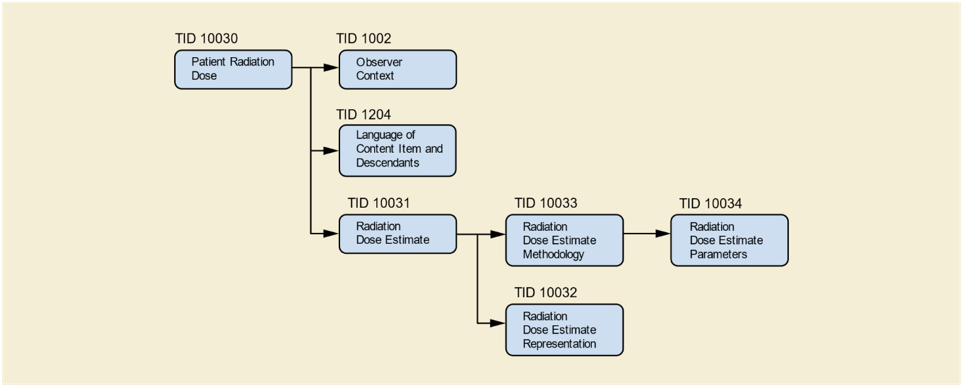
TID 10032 Radiation Dose Estimate Representation allows the dose to be stored as a separate DICOM IOD to better visualize the distribution of dose in the patient. Some examples of the representation are as a surface segmentation IOD for skin dose maps, or storing a point cloud of individual dose estimations within a patient for use in more advanced dosimetry.
TID 10033 Radiation Dose Estimate Methodology contains the information used in the dose estimation method. The first item to note is that an RDSR that stores the radiation technique factors and output is required. If the modality does not provide an RDSR, one that includes the assumed values must be created. This was done so the P-RDSR did not need to repeat the values already stored in the RDSR. The P-RDSR also provides the ability to list the values within the RDSR that were used in the dose estimation. This is needed if the dose estimation excluded some exposures, e.g., scout/localizer or setup exposures.
TID 10033 also includes the patient demographics used by the method (e.g., sex, age, weight, and height). These are only the values used in the method, and need not be the actual patient demographics. This was included to allow storage of the actual values used by the method that may be needed for unique patient habitus, etc. The TID also includes the type of model used to represent the patient (e.g., simple geometric, anthropomorphic, or actual patient segmentation models). The model can also be stored as a separate DICOM IOD, similar to the dose representation. It is hoped that this may promote the development of radiation transport and dosimetry models to be stored as DICOM IOD for standard storage and use. The registration of the patient model to the RDSR from the modality is required. Because the current RDSR does not include a frame of reference, the registration may be a simple visual registration, or it can use fiducial markers or the frame of reference from the images produced to accomplish the registration.
TID 10034 Radiation Dose Estimate Parameters is used to store any method-specific parameters used in the estimation. A listing of the parameters that may be included are specified in the DICOM Standard and the values and units for each must be provided. These includes parameters such as percent fibroglandular tissue, tissue air ratio, etc.
The detailed DICOM Standard for Structured Reports is given in Part 16 Content Mapping Resource and examples of how to use the new P-RDSR can be found in Part 17 - Explanatory Information. Physicists should learn how to create and store DICOM IOD such as RDSR and P-RDSR so they can use these standardized storage options of their work in patient dosimetry. If you have questions about the P-RDSR or any other DICOM questions please feel free to contact me, as Chair of the AAPM Working Group on DICOM Coordination and Co-Chair of MITA/DICOM WG28 Physics.
We have noticed that you have an ad blocker enabled which restricts ads served on this site.
Please disable it to continue reading AAPM Newsletter